Treatments
- Cataract Surgery
- Retina Surgery
- Glaucoma Surgery
- Pterygium Surgery
- Chalazion Surgery
- Eye Tumor Surgery
- Entropion Surgery
- Photocoagulation Laser
- YAG Laser
- Hard Lenses
- Internal Medicine Specialist
- Visual Acuity Assessment
- Eye Pressure Examination
- Fundus Photography Examination
- OCT Angiography Examination
- Gonioscopy Examination
- Visual Field Examination
- Eye USG Examination
- Colorblindness Examination
Cataract Surgery
Cataract surgery is a procedure to remove the lens of the eye and, in most cases, replace it with an artificial lens. This is done because normally, the lens of the eye is clear. A cataract causes the lens to become cloudy, which eventually affects vision

Retina Surgery
Retina surgery is a procedure inside the eye to repair a damaged or detached retina, preventing or restoring vision loss. The surgery is done under local anesthesia, so your eye is numb and you won’t feel pain. Your surgeon uses tiny instruments and a special microscope to work inside the eye. You may see occasional flashes of light during the procedure. Your pupil will be dilated beforehand to give the surgeon a clear view.

Glaucoma Surgery
The most common type of surgery for glaucoma is called trabeculectomy. It involves removing part of the eye-drainage tubes to allow fluid to drain more easily. Glaucoma surgery may be carried out under local or general anaesthetics.

Pterygium Surgery
Pterygium surgery is a procedure performed to remove noncancerous conjunctiva growths (pterygia) from the eye. The conjunctiva is the clear tissue covering the white part of the eye and the inside of the eyelids. Some cases of a pterygium produce little to no symptoms

Chalazion Surgery
Surgery to treat chalazion is an office procedure that takes about 15 to 20 minutes to perform. We inject a numbing agent into the eyelid and make a small incision in the bump. We will then drain the fluid and remove the material collected within the nodule. Typically, no stitches are required.

Eye Tumor Surgery
There are various ways to treat eye tumors, depending on the diagnosis, size and aggressiveness of the tumor, and other factors. Certain small tumors may respond to laser treatment or freezing (cryosurgery). In some instances, it is possible to remove a tumor surgically and still preserve vision. A technique advanced by Wilmer researchers fine-tunes radiation therapy for eye tumors, focusing it more precisely on the eye.

Entropion Surgery
Entropion repair is an outpatient procedure that typically only requires local anesthesia and a mild sedative. Depending on the exact needs of the patient, entropion repair may involve the removal of a small section of the eyelid or a skin graft to help reposition the eyelid.

Photocoagulation Laser
This laser treatment helps seal or shrink abnormal areas in the retina to protect your vision. Your pupil will be dilated, and we’ll use a special lens to focus the laser beam. You may see flashes of light during the brief, painless procedure. It’s a precise way to treat conditions like retinal tears or diabetic retinopathy, helping to stabilize the retina and prevent further issues.


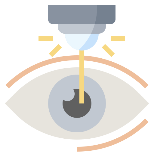
YAG Laser
The YAG laser is the laser used to clear the frosting from the back surface of an intraocular lens. YAG laser treatment is painless and is completed from outside the eye in a few minutes. During YAG laser treatment your eye doctor may use a magnifying contact lens to help with aiming the YAG laser at the layer of frosting. During the treatment patients will see flashes of light and hear a clicking sound. The pupil needs to be dilated before YAG laser can be performed to allow a good view of the lens surface.

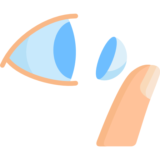
Hard Lenses
Getting fit for hard contact lenses is a specialized and precise process designed for optimal vision correction and eye health, featuring two main types: Ortho-K (Orthokeratology), which are worn overnight to gently reshape the cornea and provide clear, unaided vision during the day, and RGP (Rigid Gas Permeable) lenses, which are worn daily and renowned for providing exceptionally sharp vision, making them an ideal solution for high astigmatism or irregular corneas.


Internal Medicine Specialist
- Perioperative Evaluation Before Ophthalmic Surgery
- Consultation For all internal medicine problems
- Heart evaluation by electrocardiogram (EKG)
- Breathing therapy with nebulizer
- IV line insertion and removal
- Urinary catheter insertion and removal
- Nasogastric tube (feeding tube) insertion and removal
- Stomach cleansing (gastric lavage)
- Wounds care
- Tuberculine skin test (Mantoux test)
- Adult vaccinations service
- Joint injection for pain relief


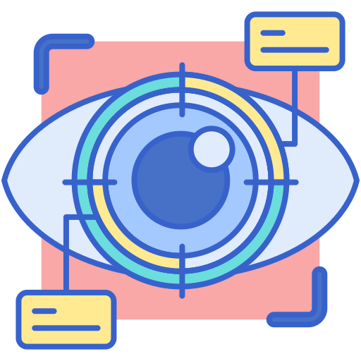
Visual Acuity Assesssment
An eye exam involves a series of tests to evaluate vision and check for eye diseases. Our doctors are likely to use various instruments, i.e. shine bright lights at the eyes and request that patients look through an array of lenses. Each test during an eye exam evaluates a different aspect of their vision or eye health.

Eye Pressure Examination
Tonometry is a quick and simple test that checks the pressure inside the eyes. The results can help our doctors see if our patient is at risk for glaucoma.


Fundus Photography Examination
Fundus photography is the process of taking serial photographs of the interior of the eye through the pupil. A fundus camera is a specialized low-power microscope attached to a camera used to examine structures such as the optic disc, retina, and lens.


OCT Angiography Examination
Optical coherence tomography (OCT) is a non-invasive imaging test. OCT uses light waves to take cross-section pictures of the retina. With OCT, our ophthalmologists can see each of the retina’s distinctive layers. This allows our ophthalmologists to map and measure their thickness.
")
Gonioscopy Examination
Gonioscopy is a painless examination to see whether the area where fluid drains out of the eye is open or closed. It is often done during a regular eye examination, depending on age and whether or not the patient is at a high risk for glaucoma.

Visual Field Examination
Humphrey visual field analyser (HVFA) is a tool for measuring the human visual field that is commonly used by optometrists, orthoptists and ophthalmologists, particularly for detecting monocular visual field.

Eye USG Examination
An eye and orbit ultrasound is a test to look at the eye area. It also measures the size and structures of the eye

Colorblindness Examination
A color vision test, also known as the Ishihara color test, measures our patient’s ability to tell the difference among colors. If they don’t pass this test, they may have poor color vision, or our doctors may tell them that they’re color blind.
